Monday, January 31, 2011

Hoping for Recovery - After the Cardiac Cath


If you've read the surgeon's perspective post or the main cath post previously, you know already that what was discovered during the cath was a big enough deal to send Lillian back to the CICU (Cardiac Intensive Care Unit) after the procedure for the long term. It is normal for kids to be sent to the CICU after a cath, but most are only there for a short while, rather than for long term management, which is why Lillian was sent back. 

Normal heart with 2 coronary arteries

With the discovery of the single coronary physiology (to recap, she only has one coronary artery on the right side of her heart, normal is one on each side of the heart, see picture above), we knew Lillian would need special care even with the coarctation (blockage) cleared. We did know from the Norwood procedure that the right coronary artery she does have mutated and expanded to go up the left side, but we weren't sure at the time how the blood flowed through her heart. The cath confirmed that there is no blood flow through the left coronary artery (perhaps its a misnomer to say she has a single coronary physiology as it is my understanding that she does in fact have a left coronary artery; but it is so small (~1mm) that there is no blood flow through it, and it is too small for us to balloon it). Even though Lillian doesn't have the pumping chamber on the left side of her heart, it still needs to be fed blood otherwise it might cause the side that she does have to die out. This then was our goal in the CICU; to see if we could either restore blood flow through the left coronary artery (it possibly had blood flow at a time before the cath, but we don't know if it did or not) or if we could get enough blood flow through the right coronary artery to sustain her heart.

To help keep Lillian's heart stable and strong after the surgery, and to hopefully accomplish either getting enough blood flow to the left side of the heart, we began Milrinone therapy when she returned to the CICU. Those of you who have been reading since the beginning might remember Milrinone as one of the medicines she was being given after the Norwood, and it serves the same purpose here; mainly dilating the coronary arteries feeding the heart making it easier to get blood to her heart, thus relaxing it and making it pump better. Milrinone also seems to have some sort of healing properties restoring some amount of function to the heart even after the medicine is stopped, although I don't entirely understand why this effect lasts in some (but not all) cases. That was what we were going for, as Lillian would need to recover heart function to continue along the surgical track, which is what we would prefer her to be on. 

The first day post cath/start of Milrinone therapy made us optimistic, Lillian's hands and feet were warm to the touch, something that we had never experienced before (as a blood mixer who doesn't have the standard 95 to 100% saturations, Lillian's extremities are typically cold). Her BNP had also gone down to 1220, a promising improvement from the 1740 reading that caused us to do the cath emergently in the first place. (as a refresher, BNP is a hormone that indicates heart distress, a normal person without heart disease should be under 100, and over 500 is generally considered catastrophic, so Lillian was high to say the least). She also was acting like she was feeling better, getting back to her happier self.


Even though Lillian was showing promising signs on the Milrinone therapy, Children's staff still wanted to have Lillian evaluated by the heart failure/transplant doctors as the single coronary physiology is not normally sustainable. The heart failure doctor (we spoke with Dr. Yuk Law, who is the director of the Heart Transplant program but one of 3 rotating doctors; the other two are Dr. Mariska Kemna and Dr. Robert Boucek; they each are attending for a week at a time, rotating between the three of them) was hopeful but not optimistic. He told us it was unlikely that Lillian would be able to remain stable with her single coronary physiology, but that we would have to wait and see how she responded to the Milrinone therapy. 

Lillian's response to Milrinone therapy was eventful to say the least. The second day after the cath Lillian's BNP shot up to 2570, the highest it had ever been and far higher than we would have expected it to be after the cath. However, even with the numbers showing alarming information, Lillian was acting fine, so we continued with the therapy. The day wasn't free from drama though. Lillian had an arterial line placed with the cath to deliver medicine, but we needed to remove it due to being unable to stop the site from oozing blood. This removal caused her blood pressure to disappear in her legs for 15 minutes, turning them blue while the nurses worked to get blood flow back. In the end, no harm was done, but it's always a little disconcerting losing pulses in any part of the body on a heart patient. 
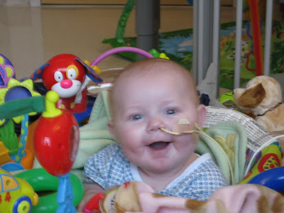
Sleeping peacefully
By Wednesday the 27th, 4 days after the cath, we still had not seen any sign of recovery in terms of heart function. Her heart was still pumping roughly the same amount of blood as before, and there were still issues with the right ventricle pumping properly. I forget if I've mentioned this before (and forgive me if I'm restating what is already in this post, this one was written over a month due to other obligations), but the right ventricle that she does have has issues with properly squeezing the bottom half of the chamber, pumping less blood than she needs and presenting the risk of a clot forming inside the heart (as another aside, if you go back to the cath posts, you can see this dysfunction in the chamber she does have) We needed to ensure proper blood flow to the heart so this chamber didn't get any weaker, as it is the only one she has. Without seeing any improvement at this point the Dr. Law already thought that she wouldn't recover enough and would have to go down the transplant path, but we weren't rushing to that decision considering it's gravity.


I'm going to jump on a little tangent here and explain the process for deciding treatment for HLHS and other congenital heart disease kids in general. Most kids with minor CHD problems are better served with appropriate surgery as their problems and and surgical side effects are relatively manageable. We also try to do surgery whenever possible for kids with more serious conditions like HLHS or coronary fistula (where the coronary arteries are inside the heart rather than outside of it); but surgery sometimes isn't enough because of the severity of the condition. For the children who have surgery but still won't survive with it, we have one final option: a heart transplant. There are advantages and disadvantages to both the surgical track and the transplant track, so many that I can't hope to list more than the most significant here. In terms of the surgical track, the advantages are that you have your own heart, and even if it is surgically repaired, your own heart is generally better than a transplanted one due to the anti-rejection medicines all transplant patients must take for the rest of their life. There is no risk of rejection with your own heart, but the risk of the repair failing at some point is always there. However, the surgical track is not fool proof. The 5 year survival rate on the first 3 surgeries of HLHS is around 70%, so they're not without risk. The first surgery, the Norwood, has a 10% fatality rate alone due to the stress of the operation. The transplant track has the disadvantage of taking anti-rejection medicines; medicines so strong that you need other medicine to counteract their side effects, and then other medicine to counteract the first other medicine's side effects (whew!). Anti rejection medicines are also immune suppressants, so taking them is tantamount to giving yourself AIDS. They have many other side effects, ranging from excessive hair growth to liver and kidney failure. At this point you're probably asking why would anyone do a transplant? The answer to that question is that once a CHD child has a transplanted heart, they no longer have CHD. The transplanted heart is a healthy, fully functional heart that allows for a normal life outside of the medicines, and we can partially manage their side effects or reduce them over time. This is in comparison with a Fontan circulation child (a child who has finished the surgical track for HLHS), who despite having their own heart, still has half a heart. They have much less energy than normal, hit brick walls in physical activity, tend to be extremely petite, and the girls can never have children due to the stress it would cause. None of these issues apply to transplant patients, they have normal hearts. Of course now you're asking why don't we do transplants for every CHD patient who needs it? And the simple answer is that we would, but we don't have enough hearts. There's not exactly an abundant supply of infant hearts (and please don't get me wrong, this is a good thing); and the small number of infants who do unfortunately pass so early either don't have their organs donated by their families for various reasons or aren't suitable donors. The lack of donors is the sole reason that the surgical track was created in the first place, with the transplant track now being reserved for the most serious of patients for whom even the surgical track will not work. 


Now that you understand the reasoning for not giving every CHD patient a transplant, you should understand a little better why we wanted to see if Lillian could recover function and stay on the surgical track. Even though we hadn't seen any sign of heart function recovery yet, we still wanted to give Lillian the best shot possible for it. With that in mind, we planned an X-ray and echocardiogram for October 31st, later pushed back to November 1st, which would determine which track we would be on. If she recovered function, we would stay on the surgical track, and if she stayed the same, we would have to go transplant.

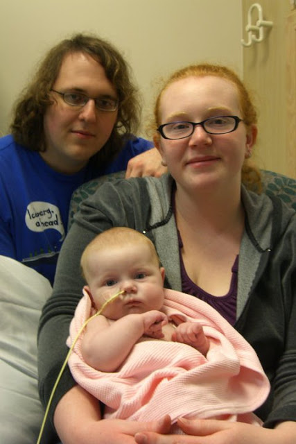
Lillian on the day of the ECHO
The ECHO results showed us exactly what we had seen earlier: no improvement at all. However, despite our understanding that this would put us on the transplant list, the heart failure team wasn't ready to commit to that option first. Even though she hadn't recovered any function, they told us what really mattered was if the function that she did have would be enough to sustain her. For some kids, a little loss in heart function isn't enough to sustain them. In contrast, other kids can lose a ton of function and still act and behave normally, eating properly and everything. In the end, this is what matters. In that regard, we decided to do a Milrinone wean, seeing how well Lillian's heart would function without it. If she couldn't function without it, we would list her and if she could, she would remain on the surgical track. Kathryn and I wanted to start the listing process at this point, as our gut feeling was that the wean would fail, but to be listed you have to demonstrate actual need. Failing a Milrinone wean is one such way to demonstrate this need, so we needed to see if she would fail or not before she was listed.


Milrinone weans are typically done as a half cut a day. That means that if you are at 0.5 mcg/kg/min as Lillian was, you would go down to 0.25 on the first day, 0.125 on the second, and then either to 0.0625 or 0, depending on the patient. We knew Lillian was fragile, so we planned on doing the wean at 0.1 mcg/kg/min a day, so that she would be off of it entirely after 5 days. Lillian reacted extremely poorly to this wean, with higher oxygen saturations on the first day, and drastic blood pressure drops and vomiting on the second, failing the wean at 0.3 mcg/kg/min. However,she had shown some positive signs during the wean, including a drastically lower BNP, as low as 200.


With Lillian failing the wean, Kathryn and I started to push heavily to start the transplant listing process. However, Children's staff wanted to do one more trial wean, but slower this time and with other possible causes removed. I'm still not sure why we weren't able to start the transplant evaluation at this point, but as the surgical track is my preferred option, I was OK with trying one more wean. This wean would still be at .1 mcg/kg/min at a time, but this time turned down every two days as opposed to every one, effectively making this wean 5 times slower as a typical one. We also switched her NG feeding tube (ends in her stomach) to a ND feeding tube (ends in her upper intestines) hoping to rule out acid reflux as a possible cause of the vomiting that caused us to abandon the wean last time. 


While waiting at the second day of the wean (still at .4 mcg/kg/min) we were able to begin the transplant listing process by drawing and completing a blood work evaluation, the single most important pre transplant evaluation. Most of the rest of the evaluations would have to wait on the results of the wean, but it was encouraging that we were at least tentatively starting them so that we could list her quickly if the wean did fail. However, the wean was looking like it was going well, so we finally had some good news. Lillian sailed through the 0.3 mcg/kg/min rate that she had failed at the first time, and she got all the way through the wean so that we could turn it off. We were hopeful that she would actually be able to stay stable with the Milrinone off, but with her BNP skyrocketing up to 700 we knew that she might have to be put back on Milrinone at any moment. But just seeing the Milrinone off was encouraging, and the fact that the ECHO looked unchanged (which is good since it wasn't worse) gave us a little more hope for getting out of the woods.


Lillian did well the first day of having the Milrinone off, but spit up a total of 10 times on the next day, a traditional sign of heart failure for her. However, her BNP was down to 319, so she was giving us very mixed signals. These signals were enough for us to continue a little further in the evaluations, doing both the cardiac anesthesia and nutrition evaluations on Lillian and a blood antibody check on Kathryn to see what she might have passed on to Lillian during birth. 

Annabelle getting in on the doctor action

At this point, even with Lillian fully off the Milrinone and stable for the moment, we decided to go full steam ahead with the transplant evaluation. Lillian had been declining slowly since we had turned it off; but we also knew that she had recovered absolutely zero heart function over the Milrinone therapy, and we knew her heart wouldn't be strong enough to sustain the circulation that the second surgery would give her. Her heart was barely able to sustain her circulation as it was. She was also spitting up like crazy and getting worse every day. We would still wait and see how she did long term off Milrinone, but in the mean time we also did her HIV evaluation (mandatory for all transplants as she had a small chance of getting it from a blood transfusion earlier), social work eval, infectious diseases eval and a neurology eval, all with the goal of being able to list her quickly if and when that decision was made.


Lillian remained fairly stable for a while after the Milrinone was turned off, but she started getting worse not long after that, spitting up more and giving us other signs. Despite the way things looked, Children's still decided to send us to floor even with Lillian's declining condition, with the expectation that if the decline continued, we would be right back in the CICU. This proved to be correct, as we were back in the CICU after 3 days; brought around by a dramatic show of 6 emesis (spit up/vomiting) in 45 min and a drop in her heart rate into the 80s (normally in the 120s to 160s).
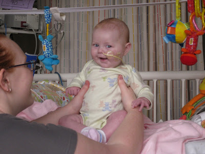
Derping it up on the floor before going back to CICU
At this point, the need for a transplant was clear and we knew that was the path we would be taking. But before Children's does any non emergent surgery or treatment, the patient's case and back story are presented at a multidisciplinary conference to get as much input and make the best choice possible. Lillian was presented at the conference for transplant on November 22nd, where the consensus was transplant, as expected, depending on one more evaluation. That evaluation, a HLA antigen eval, came back as extremely weak (which is what we want), allowing us to sign the consent form, the last step to listing. Of course, it should be the last step before transplant, but we then had to deal with a 2 day hiccup caused by a lazy Cigna case worker leaving work at 1:30PM and Cigna wanting a dental evaluation on a 4 month old baby without teeth. Why any one would need a dental evaluation for a heart transplant is beyond me, but especially when you don't even have teeth. 

Despite the road blocks that Cigna tried to throw up, Lillian was listed for a heart transplant on November 24th, 2010. She was listed as Status 1A (in order, they are 1A, 1B, 2 and 7, or inactive), and was number one on the list for her priority (the highest) in her blood type (A) in our region (region 6, encompassing Washington, Oregon, Idaho, Montana, Alaska and Hawaii) of her size. A mouthful for sure, but there are many qualifiers. The most important of these qualifiers is size, as an infant can generally receive a mismatched organ and adapt to it due to the relative immaturity of their immune systems. This means that even though Lillian is blood type A, she could receive a B heart (provided there were no other B patients of course) and generally do fine, while this would be fatal for an older child or an adult.


Not much has happened between the listing and today. We were in the hospital for Christmas, where Lillian and Annabelle got a personal visit from Santa and presents from the hospital staff.That's functionally the biggest event that happened, so I'm going to wrap up this uber long post with a quick picture dump of the 2 months between the listing and today.

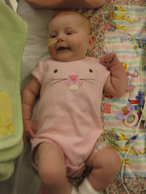
Being cute for Christmas

Asleep with our Project Linus blanket


Hamming it up

And for good measure, one of Annabelle's legendary photobombs.


It's been an interesting few months waiting for a transplant. The hospital has functionally become our home, although we still use our actual one for sleeping, if not much else. Lillian has come a long ways even from the listing, and is getting smarter and more aware every day. The similarities between her and Annabelle are really starting to show through, although I feel that Lillian will be shy in the long run due to how much of her life she has spent at the hospital. It is now January 31st. We have been in the hospital since October 22nd (102 days, or 3 months, 10 days), and listed since November 24th (69 days, or 2 months, 8 days).

Monday, January 10, 2011

The Cardiac Catheterization - The Surgeon's Perspective

Like my earlier post with the surgeon's perspective on Lillian's Norwood procedure, this post is going to be a copy of the surgeon's notes from the catheterization procedure with my comments inserted throughout [like this] to clarify. However, I do not have a medical degree, so it is possible I may have made an error in my interpretation of the notes. If I am wrong, or if I have left things uncommented when they really should be, please let me know so I can improve the post. I have previously posted the angiograms in the post before this one on the cath, but since Dr. Agustin Rubio goes into specific detail describing the angiograms I'm going to repost the videos when he is describing them to hopefully give the videos some added context. 

BACKGROUND: Lillian is a 3-month-old female who was born with complex congenital heart disease consisting of hypoplastic left heart syndrome (AAMA type). She also carries a diagnosis of a single right coronary artery [see paragraph 7 under the heading Lillian's Norwood Procedure on this post]. She has undergone a Norwood palliation and placement of a Sano shunt. Over the course of the past several weeks, she has been noted on echocardiogram to have a change in her right ventricular function as well as worsening atrioventricular valvar insufficiency [the valve is leaking, specifically, this is the tricuspid valve (between the right atrium and right ventricle), as Lillian's mitral valve is nonexistent as a side effect of the HLHS]. Her saturations continue to be rather elevated of 85-90% on room air [as Lillian is a mixer, we would want her to be 75-85% on room air]. She has been referred to the Cardiac Catheterization Laboratory due to echocardiographic findings which have not been able to adequately evaluate the distal Norwood anastomosis [we can't see the site of the tissue graft on her aorta very well]. With the evidence of decreased right ventricular function, worsening atrioventricular valvar insufficiency and elevated saturations, the suspicion is that there may be significant obstruction of the distal Norwood.

PROCEDURE/INDICATION: Right and left cardiac catheterization with possible balloon angioplasty [a balloon angioplasty is when they inflate a balloon in a blood vessel to clear a blockage] of the distal aortic arch for possible arch obstruction.

PROCEDURE: The patient's parents underwent informed consent prior to the patient coming to the Cardiac Catheterization Laboratory. The patient was brought to the Cardiac Catheterization Laboratory by Dr. Michael Richards. The patient was placed on the cardiac catheterization table and underwent appropriate sedation, analgesia [pain medicine] and securing of hemodynamic monitoring [very specific blood pressure monitoring in several different vessels].

Once the patient was sedated and adequately monitored, the patient was intubated [all infants are intubated when they are put under to reduce the risk of the lungs collapsing] and placed on room air. The patient was prepped and draped in a typical sterile fashion and 2 mL of 1% lidocaine [local anesthetic] was administered subcutaneously [i.e., a shot] into the right groin. The right femoral artery was easily accessed with a 3-French arterial catheter as was the right femoral vein and a 5-French sheath [this basically is the size of what they used] was easily placed via Seldinger technique. A 5-French wedge catheter was utilized to obtain all of the hemodynamic data throughout the right heart and left heart structures as well as into the descending aorta [basically they took measurements of all her information before doing anything else]. An 0.025 angled Glidewire was utilized to obtain a pressure beyond the distal Norwood [checking blood pressure after the location of the potential blockage]. At the time of crossing the distal arch over the wire, a severe change in right ventricular function with severe drop in blood pressure was noted. It is for that reason that the Glidecath was emergently removed and a dose of epinephrine was administered. The patient was allowed to recover prior to any further interventions occurring. [I'm not really sure why this occurred. It is possible that the Glidecath filled the vessel entirely blocking the ventricle from pumping blood and thus resulting in the blood pressure drop. I don't entirely see how this is possible though due to how small the cath is. Regardless, they gave her adrenaline, so it was enough of a concern for that relatively drastic measure.]

After the patient had her blood pressure recovered, she had 485 unites of intravenous heparin administered [a blood thinner to make sure the catheters don't cause any issues]. An angiogram [this is the X-ray videos that along with fluoroscopy allow us to have a high quality view of Lillian's blood flow] was performed via the 4 Fr angle Glide cath. A severe caliber change was noted at the distal anastomosis of the Norwood [sudden narrowing, this confirms the previously suspected blockage]. The measurement obtained revealed: 2.9mm diameter (by AP and Lat), descending aorta 5mm and 10mm (aorta proximal to arch obstruction) [this is already pretty clear, but just to emphasize, her aorta is normally between 5mm and 10mm, so only having 2.9mm open at one point is severe] The 4 Fr catheter allowed for passage of an 0.014 Hi-Torque guide-wire. The wire easily passed through the distal area of narrowing [they got through the blockage with a very thin wire]. The proximal portions of the Norwood anastomosis appeared to be widely patent as do the head and neck vessels [other than the one blockage, the rest of the Norwood tissue graft looks fine, and the vessels to her head and neck are wide open; these vessels do present something of concern in many Norwood cases, so it is good that this is open for Lillian].

The decision was made to perform a balloon angioplasty using a 6mm Tyshak II balloon dilatation catheter [this is the procedure that was speculated as being needed at the start, this will clear the blockage]. The 4 Fr Glide catheter was removed and the balloon dilation catheter was passed in prograde fashion through the narrowed segment. The balloon was quickly dilated to maximum ATM's and the[n] quickly deflated. At full inflation a very small waist [I'm not entirely sure as to what this means, but from my understanding this means that there was a tiny bit of blood flow around the balloon on the front projection. There should normally be no blood flow when the balloon is fully inflated.] was noted in the AP projection but no waist appreciated in the lateral projection. A follow-up ascending angiogram was performed which demonstrated a moderate improvement with a small degree of neointimal disruption appreciated without extravasation of contrast [the follow up angiogram shows an improvement, and although there is still a small blockage in the artery, it does nothing to the contrast, and thus will not do anything to the normal blood flow]. Improved flow of contrast through the distal transverse arch is noted with a significant improvement in the right femoral arterial pressure [the clearing is making it easier for her heart to pump blood to her extremities].

Given the significant concern for decreased myocardial function secondary to distal arch obstruction [even with clearing the blockage, they were concerned that her heart muscle would not be functioning well enough], it was felt after discussions with the cardiac intensivists and with the Heart Failure Team that Lillian would do best by being transferred to the Cardiac Intensive Care Unit with a central venous catheter and an arterial line [these allow for central medication delivery and continuous blood pressure monitoring].

Upon completion of the hemodynamic and angiographic assessment and interventional aspect of the procedure, the 5-French venous catheter was switched out for the 5-French double-lumen central venous catheter. The 3-French right femoral arterial line was kept in place.

Lillian was extubated and awoke and transferred to the Cardiac Intensive Care Unit for further medical management.

COMPLICATIONS: None.

FLUOROSCOPIC TIME: 16.7 Minutes.

CONTRAST: A total of 21 mL of Optiray contrast was administered.

I performed all aspects of the above-mentioned procedure.

RESULTS:
1. CATHETER COURSE: The catheter coursed normally through the intracardiac chambers of a hypoplastic left heart syndrome and into the left upper and right upper pulmonary veins. Distal arch was accessed in antegrade fashion [they went with the flow of blood rather than against it]
2. OXIMETRIES: The mixed venous oxygen saturation was 48% with a right atrial saturation of 69%. The systemic arterial saturation was 88% with a descending aortic saturation of 88%. The right ventricular saturation was 88%. The left upper and right upper pulmonary vein saturations were 97% with a left atrial saturation of 97%. [to clear up exactly what he's talking about: mixed venous refers to how she mixes both oxygenated and unoxygenated blood; her right atrium is the main collection chamber for blood before it is pumped, systemic refers to the part of the circulatory system that sends oxygenated blood to the body; the descending aorta delivers that oxygenated blood to the lower portions of the body; the right ventricle is her one and only pumping chamber, so should have higher saturations; pulmonary refers to the lungs so these of course should have the highest saturations; he also measures the left atrial saturation at 97%, this is expected as the left side of the heart would normally pump the oxygen rich blood, but she does not have a left ventricle to pump that blood so that chamber serves basically as a collection chamber supplementing the right one that she does have].
3. PRESSURES: The mean SVC pressure was 9mmHg. The mean right atrial pressure 9 mmHg. The mean left atrial pressure was 10 mmHg with a right ventricular pressure of 61/10. The ascending aortic pressure proximal to the area of stenosis [by the narrowing that was cleared] was 60/32 with a mean of 42 mmHg with a descending arterial pressure of 55/32 with a mean of 40 mmHg. Right and left upper pulmonary veins had mean venous pressures of 9 mmHg. The right branch pulmonary arterial pressure by pulmonary vein wedge pressure was 19/12 with a mean of 15 mmHg. The left branch pulmonary arterial pressure was estimated via pulmonary vein wedge pressure of 20/13 with a mean of 16 mmHg. Her PVR was estimated at 1.23 Woods units x m2 with a Qp/Qs of 1.13/1 [see my comment on the oximetries section for an explanation of where these are being measured; the PVR is pulmonary vascular resistance, this is what the blood must overcome to flow to the lungs. This is normally 0.25 - 1.6 Woods, but I'm unsure if that is for adults, children or both. We want this to be a little higher in Lillian to prevent too much blood from going to her lungs at the expense of going to the rest of her body].

CINEANGIOGRAMS: [I am linking the actual videos of these after he is describing each of them.]
1. Ascending aortogram: An antegrade catheter was positioned in the proximal portion of the transverse arch [the part of her aorta between the heart and the top of the arch] through the Norwood anastomosis [it was accessed through the Norwood graph rather than through the heart for obvious reasons]. Evidence of a distal arch obstruction was appreciated. The measurement at the level of the arch was 2.9 x 2.9 mm. [this is where he saw and measured the arch obstruction]. Antegrade flow into the head and neck is easily appreciated through a left-sided aortic arch [even with the blockage, we have correct flow to her head and neck]. At the postero- inferior portion of the Norwood anastomosis at the level of the native aorta, one sees an extremely hypoplastic segment. [i.e., an extremely small segment of her aorta at the portion the Norwood graft ends and her own natural aorta begins]. Antegrade flow is seen through the coronary artery into a single right coronary arterial system. No contrast is appreciable through the left anterior coronary distribution [we see normal blood flow to the arteries that supply the heart, but only through the right coronary artery; we see no blood flow through the left artery. This is by far the biggest reason the heart that Lillian does have isn't pumping strongly enough]. Multiple collateral vessels are noted to arise from the right coronary system and traverse apically [we see blood vessels splitting off from the coronary artery she does have on the right that we wouldn't normally expect, and they travel to the apex of the heart in a way we really wouldn't expect.

Here are the videos he was viewing when he was describing this section: 
From the front: 


From the side with the blockage pointed out:


]
2. Balloon angioplasty: A balloon angioplasty using a 6-mm Tyshak balloon was performed across the area of coarctation. The balloon was noted to inflate to its maximum atmospheres with only a minimal residual waist [there was only a small amount of blood flow around the balloon.

Here are the videos from this section:
From the front:


From the side:


]
3. Post-angioplasty angiogram: Once again, a selective injection at the level of proximal aorta [same location as #1] is performed with contrast noted to enter into the single coronary arterial tree and better filling the right coronary system [seeing better blood flow in the system that feeds her heart itself, yet still without any appreciable blood flow to the left side]. Lateral projection demonstrates a moderate improvement in the dimensions of the distal aspect of the transverse arch post-angioplasty. [i.e., a moderate improvement in how wide the aorta is at the site of angioplasty

Here are the videos from this section:
From the front: 


From the side:


]
4. Right ventriculogram: A right ventriculogram was performed to evaluate the Sano conduit [as a refresher, the shunt goes from her right ventricle to her pulmonary arteries to supply blood to her lungs]. There is a moderate to severe myocardial systolic contractility. [Again, I'm not exactly sure what he's referring to here. Myocardial refers to the heart muscle, systolic to the oxygen feeding circulation, so I know he is talking about there being an issue with general heart function, but he stops short of saying dysfunction, which I would expect.] Antegrade flow through the proximal, middle and distal portions of the Sano shunt appears to be widely patent [i.e., there is absolutely no issue with the shunt]. No evidence of distal branch pulmonary arterial stenosis. The branch pulmonary arteries are noted to fill in antegrade fashion with no evidence of obstruction. The distal right and left branch pulmonary arteries appear to be of adequate caliber. [The last 3 sentences basically say there's no evidence of a narrowing in her pulmonary arteries, which again go to the lungs; and there is normal blood flow.

Here are the videos from this section:
From the front:


From the side:


]
5. Final angiogram: A final angiogram using a Berman angiographic catheter was performed in the transverse arch. This demonstrates improved caliber across the distal Norwood anastomosis with improved filling of the single right coronary artery. Despite the catheter across the neo-aortic valve, no evidence of neoaortic valvar insufficiency is appreciated. On the lateral projection, a previously unnoticed region at the distal aortic arch reveals a neointimal disruption with no extravasation of contrast. [He saw a new small blockage in the arch, but it is not causing any issues.

Here are the videos from this section:
From the front:


From the side: 


]
IMPRESSION: Lillian is a 3-month old with complex congenital heart disease consistent of a severely hypoplastic ascending aorta, and mitral atresia [there is no valve between the two chambers on the left side of the heart, this is insignificant as she doesn't even have a left ventricle]. She has undergone a Norwood palliation for her hypoplastic left heart syndrome with the inclusion of a Sano shunt. The Sano appears to be widely patent with antegrade flow noted into the right and left branch pulmonary arteries [this is as expected]. No evidence of pulmonary vein abnormalities is noted. She demonstrated a severe coarctation of her distal arch which underwent balloon angioplasty and resulted in an improved flow by contrast injection [coarctation = blockage, this is why she needed the cath]. She was to be transferred to the Cardiac Intensive Care Unit for systemic afterload reduction [giving her heart a rest] and acute management of her heart failure. A double lumen central venous catheter replaced her venous sheath in her right femoral vein and the single lumen arterial catheter was placed into the right femoral artery. These catheters will remain in place so as to allow for systemic medications to be delivered [they also allow us to draw labs without poking her]. Her data and information will be presented at the next patient care conference.

Sunday, January 2, 2011

The Cardiac Catheterization - Trying to Save Lillian's Heart


Like the Norwood surgery post, this post is intended primarily to explain what a cardiac catheterization is in general terms as well as explain how it went for Lillian. There will be a follow up post with the surgeon's notes later. As before, this will be much easier to follow if you've familiarized yourself with Lillian's heart condition, which you can read about here, as well as (to a lesser extent) her first surgery, which you can read about here.

A cardiac catheterization is a minimally invasive surgical procedure that allows the catherterization doctor/surgeon very clear views of the patient's heart and blood flow; in addition to allowing for other procedures like angioplasties. The way they are able to view the heart is by taking special X-rays called angiograms that allow extremely clear images of the heart. One such image is seen above. The procedure starts with surgeon inserting a guide wire into either the femoral artery (in the groin) or the radial artery (in the wrist). The surgeon then guides a plastic sheath over the guide wire and removes the guide wire once the sheath is in place. This sheath allows for the insertion of the cathether which is guided through the artery to the heart using another guide wire and X-rays. Once it reaches the intended location, the surgeon can inject special dye that is visible to X-rays or perform other procedures.

Lillian's cardiac catheterization was performed by Dr. Agustin Rubio (who really needs a higher resolution picture on Children's webpage...) on October 23rd. Since Lillian's veins and arteries are so small they used her left femoral artery as the entry point. Once the catheter was at her heart, Dr. Rubio performed a dye injection in her aorta to assess the state of her heart. One cool thing about the cath though is that it produces great pictures/videos. You can see the videos from this dye injection below.

A couple of notes about the videos - Youtube has compressed the picture quality so some of the really amazing detail is missing. If you're interested in seeing the videos in more detail, I have the full copies on my computer, and am willing to show them to other people. Regardless, I recommend watching it in the highest quality possible. The high quality video where you can see the dye flow in not only the major veins but the small ones as well is quite amazing. Also, the things you are seeing in the videos: the loops (there are 5 or 6 visible) are her stitches under her skin that keep her chest closed. The spring looking object (I believe) is the Sano shunt, and the two black lines I think are bands around the pulmonary arteries.

Start of Cath Dye Injection - Aorta - Front Profile 

Start of Cath Dye Injection - Aorta - Side Profile.

This dye injection revealed a severe coarctation (blockage) of Lillian's aorta near the end of where it tissue graft ended caused by scar tissue from the surgery. Even with the repair and expansion of the aorta done as good as it could be done, scar tissue is still an occurrence in about 15% of patients who get a Norwood procedure. The scar tissue left only an opening of 2.9mm in diameter, when her normal aorta ranged from 5mm to 10mm. Pumping blood past this blockage was likely the cause of the heart distress, so the doctors decided to perform a balloon angioplasty of the blockage to remove it. 

Aortic Angioplasty - Front Profile

Aortic Angioplasty - Side Profile

The blockage in the aorta was pretty severe, and Dr. Rubio was only able to do one angioplasty. Even that angioplasty caused her to code briefly, and it was not worth the risk to do a second one to be sure. Once the angioplasty was completed, Dr. Rubio and his team did more dye injections to assess function after the blockage was cleared. The videos for those injections are below. 
Post Angioplasty Aortic Dye Injection - Front Profile

Post Angioplasty Aortic Dye Injection - Side Profile 

These next two videos are probably the most interesting ones. This dye injection was directly into Lillian's right ventricle, the one pumping chamber she has in her heart. This video really shows the branching veins of her circulatory system; but more importantly how poor her heart function is. You'll notice in these videos that Lillian's heart takes quite a few beats to clear the dye. 

Post Angioplasty Ventricle Dye Injection - Front Profile

Post Angioplasty Ventricle Dye Injection - Side Profile

At this point another dye injection was done to see how the blood flow looked a little while loner after the angioplasty. These two videos are strikingly similar to the other two videos labeled as Aortic Dye Injections, but these videos are so interesting that I'm including all of them. 

Post Angioplasty Aortic Dye Injection #2 - Front Profile

Post Angioplasty Aortic Dye Injection #2 - Side Profile

At this point the cath was finished so Lillian was woken up and sent to the Cardiac Intensive Care Unit for care. The cath was an extremely valuable procedure to do as it told us things about Lillian's heart that we had never been able to see or tell before, even during open heart surgery. The cath showed us the blockage of her aorta that we had never been able to see via ECHO. The blockage was so bad and her heart was in such distress that she probably would have not made it through the weekend without the angioplasty.

Perhaps the most significant thing the cath has told us, or at least confirmed, is that Lillian has a single coronary physiology. In English: we all have two coronary arteries which are the blood vessels that feed the heart directly. Lillian has only one coronary artery that we can see. Technically, she does have a second one, but we see no blood flow through this so it's existence is negligible. The one she does have is small and mutated, traveling down the right side of the heart as we would expect before branching and traveling back up the left side, which we would not expect. And even though Lillian doesn't use the left side of her heart, it is still a living muscle and needs to be given blood. Without the heart getting enough blood itself, it is unable to pump enough blood to the rest of Lillian's body. This is the real cause of the symptom that caused us to notice the heart failure; as since her stomach wasn't getting enough blood, it wasn't able to digest food properly and Lillian didn't eat. 

Our hope at this point was that with Milrinone therapy, Lillian would recover enough heart function to continue along the surgical track, but that insight would only come with time.